
|
|
Panosteitis
By Daniel A. Degner, DVM, Diplomate ACVS
Return to QuailRidge Health & Training Page -
Signalment
| Young large breed dogs that are 6 to 18 months old | |||||||||||||
Common breeds affected
|
Synonyms
| Eosinophilic panosteitis | |
| Enostosis | |
| Endosteal proliferation of new bone | |
| Eopan |
Clinical Presentation
Affects the shaft of long bones
| |||||||||||
Lameness is frequently of sudden onset
| |||||||||||
| The affected bone is painful to touch | |||||||||||
Some dogs can show signs of
|
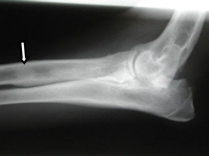
Signs on Radiographs (x-rays)
| If the disease is early in its course no abnormalities may not be seen on the radiographs; if the radiographs are repeated in 2 weeks the problem usually can be seen | |
| Increased density in the marrow cavity of the affected bone can be seen | |
| The wall of the bone becomes thicker due to new bone formation on the inner and outer layer of the bone | |
| Two to three months later the bone normalizes and the bone looks normal on radiographs again | |
| In the radiograph below take note of the grey spot in the marrow cavity (denoted by the arrow) which is eopan |
Microscopic Signs
| There usually is no inflammatory component of panosteitis | |
| The main change seen is fibrosis of the marrow (scar tissue develops) | |
| With time the fibrous tissue changes into bone, hence the increased density as seen on the radiographs | |
| New bone formation on the inner and outer part of the bone can be seen |
Cause
| Unknown | |
| Potentially an unidentified viral infection |
Treatment
| Self-limiting disease that has a spontaneous recovery | |
| Repeated bouts of this disease are about one month apart | |
| Problem usually does not recur after 12 to 18 months of age | |
| Treatment is supportive | |
| Anti-inflammatory medications | |
| If the pet is systemically ill, then intravenous fluid therapy may be needed for rehydration |
All illustrations, photographs and text are Copyright © 2003 Vet Surgery Central
A Secondary Source of Information on Panosteitis follows:
Panosteitis
Panosteitis is a spontaneously occurring lameness that usually occurs
in large breed dogs. German Shepherds seems to be particularly predisposed
to this condition. Due to this, it is possible that the disease may have
genetic causes. Some veterinarians feel that this disease may be induced
or worsened by stress.
Affected dogs are usually in the 5 to 14 month age range and male dogs
are more commonly infected than female dogs. The disease has been reported
in dogs as young as 2 months and can occur in young mature dogs. The lameness
tends to occur very suddenly, usually without a history of trauma or excessive
exercise. In most cases one or the other front leg is affected first and
then the problem tends to move around, making it appear that the lameness
is shifting from leg to leg. There are often periods of improvement and
worsening of the symptoms in a cyclic manner. This makes evaluation of
treatment difficult since many dogs will spontaneously recover with or
without treatment and then relapse.
X-rays usually reveal that the bones have greater density than is normally
found. If pressure is applied over the long bones, pain is usually present.
The X-ray signs do not always match the clinical signs.
In most cases, the worst pain lasts between one and two months but may
persist in a cyclic nature for up to a year. Analgesic medications like
aspirin can be be helpful. In severe cases, corticosteroids may provide
relief.
Currently, a common rumor is that low protein, low calcium diets may
prevent this condition. It should be noted that the energy level of low
protein/calcium diets is often lower as well. If this is the case, a puppy
will eat much more of the diet in order to meet its energy needs, resulting
in higher total calcium consumption. It may be preferable to feed a puppy
diet and restrict total quantity to keep the dog lean than to use a low
protein/low calcium adult dog food.
This condition is self limiting, meaning that it will eventually go
away, with or without treatment. Pain control can go a long way towards
helping your pet feel more comfortable and should be used, though.
The above information is authored by Dr Michael Richards, DVM and produced by TierCom, Inc.
Opinions expressed are those of Dr. Richards.